Digital Radiography

Digital Radiography: Transforming Medical Imaging

Digital Radiography (DR) is an imaging technique that uses digital detectors to capture X-ray images of the human body. It offers faster image acquisition, enhanced image quality, and lower radiation exposure compared to traditional film-based radiography, crucial for accurate medical diagnostics and treatment plan.
3D Image from Digital Radiography

Overview

Digital Radiography (DR) has emerged as a transformative technology in the field of medical imaging, revolutionizing the way healthcare professionals capture, process, and interpret radiographic images. In contrast to traditional film-based radiography, digital radiography employs advanced digital sensors to acquire and store images, offering numerous advantages in terms of efficiency, image quality, and accessibility. This article by Academic Block examines the intricacies of digital radiography, exploring its evolution, underlying technology, applications, benefits, and future prospects.

Evolution of Radiography

Traditional radiography, also known as analog or film-based radiography, has been a cornerstone of medical imaging since Wilhelm Conrad Roentgen's discovery of X-rays in 1895. Early radiographs were captured on light-sensitive film, a process that involved exposing the film to X-rays, developing the film using chemical solutions, and producing hard-copy images. While this technique served the medical community well for many decades, it had its limitations, including the time-consuming film processing, storage challenges, and the need for repeated exposures to achieve optimal images.

The advent of computed radiography (CR) in the late 20th century marked a significant leap forward. CR introduced the use of photostimulable phosphor plates that could capture X-ray images and then be scanned to produce digital images. This eliminated the need for traditional film and allowed for quicker image acquisition and digital storage. However, CR still had some inherent limitations, such as lower image quality compared to digital radiography and the requirement for a dedicated scanner.

Digital Radiography Technology

Digital radiography builds upon the principles of computed radiography but takes the process a step further by directly capturing and processing X-ray images digitally. There are two main types of digital radiography systems: Direct Digital Radiography (DDR) and Indirect Digital Radiography (IDR).

Direct Digital Radiography (DDR)

DDR systems utilize a flat-panel detector (FPD) to directly capture X-ray images. These detectors are composed of a scintillator material that converts X-rays into visible light, which is then detected by an array of photodiodes. The resulting electrical signals are converted into digital images in real-time, eliminating the need for additional image processing steps. DDR offers high spatial resolution and faster image acquisition times compared to traditional film or CR.

Indirect Digital Radiography (IDR)

IDR systems, on the other hand, use a two-step process to capture and convert X-ray images into digital format. First, X-rays are absorbed by a scintillator material, which then emits light. This light is captured by a photosensitive array, typically a charge-coupled device (CCD) or a complementary metal-oxide-semiconductor (CMOS) sensor. The electrical signals generated by the photosensitive array are then converted into digital images. While IDR systems may have slightly lower spatial resolution than DDR, they often provide better detective quantum efficiency (DQE), resulting in superior image quality at lower radiation doses.

Applications of Digital Radiography

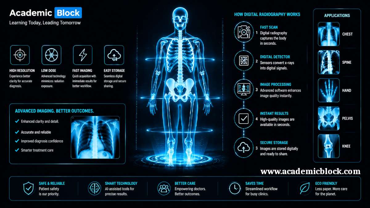
Digital radiography has found applications in various medical fields, contributing to improved diagnostics, treatment planning, and patient care. The versatility and efficiency of digital radiography systems have led to their adoption in the following areas:

  1. General Radiography: Digital radiography has become the standard for general radiographic examinations, including chest X-rays, skeletal imaging, and abdominal studies. The real-time image acquisition and immediate availability of digital images enhance the diagnostic process, allowing healthcare professionals to make faster and more accurate assessments.

  2. Fluoroscopy: Fluoroscopy involves real-time imaging of moving structures within the body, often used for procedures such as barium studies, angiography, and joint injections. Digital fluoroscopy systems provide dynamic, high-resolution images with reduced radiation exposure, benefiting both patients and healthcare providers.

  3. Dental Radiography: Digital radiography has gained popularity in dentistry for intraoral and extraoral imaging. The ability to capture and store digital dental images facilitates better collaboration between dental professionals and allows for efficient comparison of current and past images for treatment planning.

  4. Mammography: Digital mammography has become the gold standard for breast cancer screening. Digital mammography systems offer improved image quality, enhanced image manipulation capabilities, and the ability to store and transmit images electronically, streamlining the screening and diagnostic processes.

  5. Orthopedic Imaging: In orthopedics, digital radiography is widely used for evaluating fractures, joint conditions, and musculoskeletal disorders. The high spatial resolution of digital images aids in precise diagnosis and treatment planning for orthopedic patients.

Benefits of Digital Radiography

The adoption of digital radiography has brought about a multitude of benefits for both healthcare providers and patients. Some of the key advantages include:

  1. Improved Image Quality: Digital radiography systems produce high-resolution images with excellent contrast, allowing for enhanced visualization of anatomical structures. This superior image quality contributes to more accurate diagnoses and better patient outcomes.

  2. Reduced Radiation Exposure: Digital radiography often requires lower radiation doses to achieve comparable or even superior image quality compared to traditional film-based methods. This reduction in radiation exposure is particularly significant for patients undergoing repeated imaging studies.

  3. Increased Efficiency: Digital radiography streamlines the entire imaging process, from image acquisition to interpretation. The elimination of film processing and the immediate availability of digital images enable faster workflows, reducing patient waiting times and improving overall efficiency in healthcare facilities.

  4. Electronic Image Storage and Retrieval: Digital radiography allows for the electronic storage of patient images, eliminating the need for physical film archives. This not only saves physical storage space but also facilitates easy retrieval and sharing of images among healthcare professionals, leading to more collaborative and integrated patient care.

  5. Image Manipulation and Enhancement: Digital radiography enables healthcare professionals to manipulate and enhance images for better visualization of specific structures or pathologies. This includes adjusting contrast, brightness, and zooming in on specific areas of interest, providing valuable insights for diagnosis and treatment planning.

  6. Enhanced Connectivity: Digital radiography systems can be integrated into healthcare information systems (HIS) and picture archiving and communication systems (PACS), promoting seamless communication and collaboration between different departments and healthcare facilities. This connectivity contributes to a more comprehensive and coordinated approach to patient care.

Mathematical equations behind the Digital Radiography

The mathematical equations behind Digital Radiography (DR) involve the principles of X-ray physics, image formation, and digital image processing. Here, I'll provide an overview of some fundamental equations and concepts related to digital radiography:

  1. X-ray Attenuation: The basic principle behind radiography is the attenuation of X-rays as they pass through different tissues. The X-ray attenuation can be described using the Lambert-Beer law, which relates the intensity of the X-ray beam (I) before and after passing through a material:

    I = I0⋅e−μ⋅x ; where:

    • I0 is the initial intensity of the X-ray beam,

    • I is the intensity after passing through the material,

    • μ is the linear attenuation coefficient of the material, and

    • x is the thickness of the material.

  2. Image Formation - Analog System (Film Radiography): In traditional film radiography, the film response (optical density, OD) is related to the X-ray exposure (fluence) by the Hurter-Driffield curve:

    OD = −log⁡10 (E) ; where:

    • OD is the optical density,

    • E is the X-ray exposure.

  3. Image Formation - Computed Radiography (CR): In CR, a photostimulable phosphor plate is used to capture X-rays. The number of emitted light photons (N) is related to the incident X-ray exposure (E) and the conversion efficiency (CE) of the phosphor material:

    N = CE⋅E ;

    The emitted light is then detected and converted into a digital signal.

  4. Image Formation - Direct Digital Radiography (DDR) and Indirect Digital Radiography (IDR): DDR and IDR use flat-panel detectors (FPDs) to directly or indirectly capture X-ray images. The digital image formation involves the conversion of X-ray energy into electrical signals. The relationship between the incident X-ray intensity (I), the exposure (fluence), and the pixel value in the digital image (P) can be expressed as:

    P = k / I ;

    where k is a constant related to detector sensitivity.

  5. Quantum Efficiency (QE): Quantum efficiency is a measure of how efficiently a detector converts incident X-ray photons into a usable signal. It is defined as the ratio of the number of X-ray photons detected to the number of X-ray photons incident on the detector.

    QE = Number of X-ray photons detected / Number of X-ray photons on the detector ;

  6. Signal-to-Noise Ratio (SNR): SNR is a crucial parameter in digital radiography, representing the ratio of the signal strength to the noise level. In digital imaging, a higher SNR is desirable for better image quality. It can be calculated as:

    SNR = Mean signal intensity / Standard deviation of noise ;

These equations provide a glimpse into the mathematical foundations of digital radiography. The field involves complex algorithms for image processing, noise reduction, and enhancement, often utilizing techniques from signal processing and linear algebra to produce high-quality medical images.

Challenges and Considerations

While digital radiography offers numerous advantages, its adoption and implementation come with certain challenges and considerations:

  1. Initial Cost: The upfront cost of acquiring and installing digital radiography systems can be a significant investment for healthcare facilities. However, the long-term benefits, including increased efficiency and improved patient care, often outweigh the initial expenses.

  2. Training and Workflow Integration: Healthcare professionals need training to effectively use digital radiography systems and integrate them into existing workflows. Transitioning from film-based to digital processes may require adjustments in protocols and practices to optimize the benefits of the technology.

  3. Maintenance and Upgrades: Digital radiography systems require regular maintenance to ensure optimal performance. Additionally, technology evolves, and upgrades may be necessary to stay current with advancements in imaging technology and standards.

  4. Data Security and Privacy: The electronic storage and transmission of patient images raise concerns about data security and privacy. Healthcare facilities must implement robust cybersecurity measures to protect patient information and comply with relevant regulations.

  5. Radiation Dose Management: While digital radiography often allows for lower radiation doses, proper dose management is crucial. Healthcare providers must implement strategies to optimize radiation exposure, especially for pediatric and sensitive patient populations.

Future Trends in Digital Radiography

The field of digital radiography continues to evolve, with ongoing research and development leading to innovative advancements. Some emerging trends and future developments include:

  1. Artificial Intelligence (AI) Integration: The integration of AI into digital radiography is poised to revolutionize image interpretation and diagnosis. AI algorithms can assist healthcare professionals in detecting abnormalities, automating repetitive tasks, and providing decision support, ultimately improving diagnostic accuracy and efficiency.

  2. 3D Imaging and Tomosynthesis: Three-dimensional (3D) imaging and tomosynthesis are gaining prominence in digital radiography. These technologies offer enhanced visualization of complex anatomical structures and are particularly valuable in areas such as breast imaging and orthopedics.

  3. Portable and Point-of-Care Systems: Advancements in miniaturized digital radiography systems enable portable and point-of-care imaging solutions. These systems are particularly beneficial in emergency settings, intensive care units, and remote healthcare environments, where immediate imaging is essential.

  4. Advanced Detectors and Sensors: Ongoing research focuses on developing advanced detectors and sensors for digital radiography, aiming to improve image quality, enhance sensitivity, and reduce radiation doses further.

  5. Interoperability and Standardization: Efforts to improve interoperability and standardization across different imaging modalities and healthcare information systems continue. This ensures seamless integration and communication between various components of the healthcare infrastructure.

Final Words

In this article by Academic Block we have seen that, Digital radiography stands as a transformative force in the field of medical imaging, offering numerous advantages in terms of image quality, efficiency, and patient care. The evolution from traditional film-based radiography to advanced digital systems has paved the way for enhanced diagnostic capabilities and streamlined workflows. As technology continues to advance, the integration of artificial intelligence, 3D imaging, and portable systems holds the promise of further improving the precision and accessibility of medical imaging.

Healthcare professionals and institutions must navigate the challenges associated with adopting digital radiography, including initial costs, training requirements, and data security considerations. By addressing these challenges and staying abreast of emerging trends, the healthcare industry can leverage the full potential of digital radiography to provide superior diagnostic and therapeutic services, ultimately improving patient outcomes and advancing the field of medical imaging into the future. Please provide your comments below, it will help us in improving this article. Thanks for reading!

This Article will answer your questions like:

+ What is computerized radiography? >

Computerized radiography (CR) is a digital imaging technique that uses photostimulable phosphor plates to capture x-ray images. Unlike traditional film-based radiography, CR systems scan the phosphor plates after exposure to x-rays to produce digital images. These digital images can then be enhanced, stored, and shared electronically. CR eliminates the need for chemical processing of films and provides more flexible image manipulation, offering advantages in image quality, storage, and retrieval. It serves as a bridge between conventional radiography and fully digital radiology systems.

+ What is the meaning of digital radiology? >

Digital radiology refers to the use of digital detectors to capture x-ray images directly, bypassing the need for film or photostimulable plates. Digital radiography systems convert x-ray energy into electronic data that can be displayed, processed, and stored on computers. The advantages include faster image acquisition, improved image quality, reduced radiation exposure, and more efficient storage and sharing of images. Digital radiology is widely used in medical diagnostics, providing real-time access to high-resolution images and facilitating more precise diagnosis and treatment planning.

+ What is dynamic digital radiography? >

Dynamic digital radiography (DDR) is an advanced imaging technology that combines real-time motion capture with digital x-ray imaging. Unlike conventional static radiography, DDR enables the visualization of moving anatomical structures, such as the respiratory system or musculoskeletal movements, by acquiring multiple frames over time. This technique is particularly useful for functional assessments and enhances diagnostic capabilities by allowing physicians to observe physiological changes in motion. DDR is often used in pulmonary, orthopedic, and cardiovascular studies to provide both structural and dynamic information in a single imaging session.

+ How does digital radiography work? >

Digital radiography works by exposing a patient to X-rays, which pass through the body and are captured by digital detectors. These detectors convert X-ray photons into electrical signals, which are then processed digitally to create detailed images that can be viewed on a computer screen.

+ What are the main components of a digital radiography system? >

A digital radiography system includes X-ray tubes for generating radiation, digital detectors (such as flat-panel detectors or computed radiography plates), imaging processing units, and display monitors. Image acquisition software and storage systems are also integral components.

+ How does digital radiography differ from traditional film-based radiography? >

Digital radiography differs from traditional film-based radiography by eliminating the need for chemical processing of X-ray films. It offers immediate image acquisition, enhanced image quality, efficient storage and retrieval of images, and the ability to digitally enhance and analyze images.

+ What are the primary applications of digital radiography in healthcare? >

Digital radiography is used for diagnosing fractures, detecting diseases like pneumonia and cancer, assessing dental conditions, and guiding surgical procedures. It is essential in emergency medicine, orthopedics, dentistry, and general medical diagnostics.

+ What are the advantages of using digital radiography over conventional methods? >

Digital radiography offers advantages such as immediate image availability, lower radiation exposure, enhanced image quality with adjustable contrast and zoom capabilities, and easier image storage and retrieval. It also supports telemedicine and facilitates rapid diagnosis and treatment planning.

+ What is digital dental x ray? >

Digital dental x-ray is a diagnostic imaging technique used in dentistry to capture high-resolution images of teeth, gums, and surrounding bone structures. Instead of traditional film, digital sensors or phosphor plates are used to capture the x-rays, which are then converted into electronic data. Digital dental x-rays offer several advantages, including lower radiation exposure, faster image acquisition, and enhanced image quality. These images can be easily manipulated for better diagnosis, stored electronically, and shared with specialists, facilitating improved dental care and treatment planning.

+ What is Industrial digital x ray used for? >

Industrial digital x-ray is used for non-destructive testing (NDT) of materials and components in industries such as aerospace, automotive, and manufacturing. This technology helps inspect the internal structure of objects without causing damage, making it ideal for detecting flaws, cracks, or other defects in welds, castings, and composite materials. Digital x-rays offer real-time imaging, high-resolution visualization, and enhanced image manipulation, enabling precise analysis of material integrity. Industrial digital x-ray is critical for quality control, safety inspections, and ensuring product reliability in industrial applications.

+ How is image quality in digital radiography assessed and improved? >

Image quality in digital radiography is assessed through criteria such as spatial resolution, contrast resolution, noise levels, and artifacts. It is improved by optimizing exposure parameters, using advanced digital detectors, and employing image processing techniques for noise reduction and enhancement of diagnostic features.

+ What role does image processing software play in digital radiography? >

Image processing software in digital radiography enhances image quality by adjusting brightness and contrast, reducing noise, and improving spatial resolution. It enables radiologists to manipulate images for better visualization of anatomical details and abnormalities, aiding accurate diagnosis and treatment planning.

+ How is patient radiation exposure managed in digital radiography? >

Patient radiation exposure in digital radiography is managed through optimized exposure protocols, dose monitoring systems, and use of high-sensitivity detectors that require lower radiation doses for image acquisition. Continuous advancements in technology aim to minimize radiation while maintaining diagnostic image quality.

+ What are the limitations and challenges of digital radiography? >

Limitations of digital radiography include initial setup costs, potential for artifacts from digital detectors, and the need for periodic software updates and maintenance. Challenges include ensuring consistent image quality across different equipment manufacturers and managing large volumes of digital image data.

+ How does digital radiography contribute to faster diagnosis and treatment planning? >

Digital radiography enables rapid image acquisition, immediate availability of images for interpretation, and seamless integration with electronic medical records (EMRs). This facilitates faster diagnosis, enhances communication between healthcare providers, and supports timely treatment decisions.

+ What recent advancements have been made in digital radiography technology? >

Recent advancements in digital radiography technology include development of wireless and portable X-ray detectors, improved image resolution and sensitivity, implementation of artificial intelligence for image interpretation, and integration of 3D imaging capabilities for enhanced diagnostic accuracy in complex cases.

List the hardware and software required for Digital Radiography

Hardware Components:

  1. X-ray Source: X-ray tube: Generates X-rays for imaging. Modern systems may use rotating anode X-ray tubes for improved performance.
  2. Detector:
    • Direct Digital Radiography (DDR): Flat-panel detectors (FPDs) made of amorphous selenium or other materials.
    • Indirect Digital Radiography (IDR): Scintillator materials coupled with detectors, such as charge-coupled devices (CCDs) or complementary metal-oxide-semiconductor (CMOS) sensors.
  3. Collimator: Limits the X-ray beam to the area of interest, reducing unnecessary radiation exposure.
  4. Positioning and Support Systems: Radiographic tables and stands for positioning patients and X-ray equipment.
  5. Image Acquisition Device: If using computed radiography (CR), a device for reading and processing photostimulable phosphor plates.
  6. Control Console: Interface for technologists to control X-ray exposure parameters, view images, and manage the imaging process.
  7. Digital Image Display: High-resolution monitors for reviewing and interpreting digital radiographic images.
  8. Power Supply and Cooling Systems: Electrical systems and cooling mechanisms to support continuous and safe operation of X-ray equipment.

Software Components:

  1. Acquisition Software:
    • Controls the X-ray exposure parameters.
    • Manages image acquisition and processing.
  2. Image Processing Software:
    • Enhances and optimizes the acquired digital images.
    • Includes algorithms for contrast adjustment, noise reduction, and image sharpening.
  3. Image Storage and Retrieval:
    • Picture Archiving and Communication System (PACS): Manages the storage, retrieval, and distribution of digital images within a healthcare facility.
  4. DICOM (Digital Imaging and Communications in Medicine): Standard for transmitting, storing, and sharing medical images and related information.
  5. Radiology Information System (RIS): Manages patient scheduling, examination information, and other administrative aspects of radiology workflows.
  6. Electronic Health Record (EHR) Integration: Integration with broader healthcare information systems to facilitate seamless sharing of patient data and imaging results.
  7. Quality Assurance Software:
    • Monitors and ensures the quality of digital radiographic images.
    • May include tools for dose monitoring and management.
  8. Viewer Software: Allows radiologists and other healthcare professionals to view, analyze, and interpret digital radiographic images.
  9. Post-Processing Tools: Tools for annotations, measurements, and other post-processing tasks to assist in diagnosis.
  10. Security and Compliance Software: Ensures compliance with data security regulations and standards, protecting patient information and images.

List Key Discoveries where Digital Radiography is used

Medical Imaging:

  • Cancer Detection and Diagnosis: Digital mammography and tomosynthesis have improved the early detection of breast cancer, contributing to better outcomes for patients.
  • Cardiovascular Imaging: Digital radiography is instrumental in angiography and vascular studies, aiding in the diagnosis and treatment of cardiovascular diseases.
  • Orthopedics: Digital radiography is widely used for detecting fractures, joint conditions, and musculoskeletal disorders, providing detailed images for orthopedic assessments.
  • Emergency Medicine: In emergency settings, digital radiography allows for rapid imaging, aiding in the diagnosis and treatment of trauma and acute medical conditions.

Dental Imaging:

  • Digital radiography has transformed dental diagnostics, allowing for precise and efficient imaging in intraoral and extraoral examinations.

Industrial Testing and Non-Destructive Evaluation:

  • Aerospace Engineering: Digital radiography is used to inspect critical components of aircraft and spacecraft, ensuring structural integrity and safety.
  • Automotive Industry: Digital radiography is employed for inspecting welds, casting, and other components in the automotive manufacturing process, contributing to quality control.
  • Pipeline Inspection: In the oil and gas industry, digital radiography is used to inspect pipelines and detect defects without disrupting operations.
  • Welding Inspections: Digital radiography is crucial for non-destructive testing of welds in various industries, including construction and manufacturing.

Art and Cultural Heritage Preservation:

  • Digital radiography is employed in the examination and conservation of art and cultural artifacts, revealing hidden details and detecting structural issues without physical intervention.

Scientific Research:

  • Paleontology: Digital radiography is used in paleontological research to study fossils without damaging delicate specimens, providing insights into evolutionary history.
  • Material Science: Researchers use digital radiography to study the internal structures of materials, contributing to advancements in material science and engineering.
  • Archaeology: Digital radiography aids in the examination of archaeological artifacts, allowing researchers to analyze the internal structures of objects without physically disrupting them.

Veterinary Medicine:

  • Digital radiography is widely utilized in veterinary medicine for diagnostic imaging in animals, including small and large animals.

Forensic Science:

  • Digital radiography assists forensic experts in analyzing skeletal remains and identifying trauma or pathologies, contributing to forensic investigations.

Environmental Monitoring:

  • Digital radiography is applied in environmental science for studying soil profiles, plant root structures, and other subsurface features without excavation.

Facts on Digital Radiography

Evolution from Analog to Digital: Digital radiography represents a significant evolution from traditional analog or film-based radiography. This transition has led to improved image quality, efficiency, and accessibility in medical imaging.

Discovery of X-rays: The foundation of radiography, including digital radiography, can be traced back to the discovery of X-rays by Wilhelm Conrad Roentgen in 1895. Roentgen’s groundbreaking work laid the groundwork for the entire field of medical imaging.

Computed Radiography (CR) Precursor: Computed radiography (CR) served as an intermediate step between traditional film radiography and fully digital systems. CR introduced the use of photostimulable phosphor plates, allowing for the digital capture of X-ray images.

Direct and Indirect Digital Radiography: Digital radiography is categorized into two main types: Direct Digital Radiography (DDR), which uses a flat-panel detector (FPD) to directly capture X-rays, and Indirect Digital Radiography (IDR), which involves a two-step process using a scintillator and detector.

Flat-Panel Detectors (FPDs): FPDs are a key component of digital radiography systems. These detectors, made of materials like amorphous selenium or cesium iodide, directly convert X-rays into electronic signals, enabling the creation of digital images.

Advantages of Digital Radiography:

Improved Image Quality: Digital radiography provides high-resolution images with excellent contrast.

Reduced Radiation Exposure: Digital systems often require lower radiation doses compared to traditional film.

Increased Efficiency: Immediate availability of digital images streamlines workflows.

Electronic Image Storage: Digital images can be stored electronically, facilitating easy retrieval and sharing.

Artificial Intelligence (AI) Integration: The integration of AI algorithms in digital radiography is a growing trend. AI enhances image analysis, assists in diagnosis, and supports decision-making for healthcare professionals.

3D Imaging and Tomosynthesis: Digital radiography has evolved to include three-dimensional (3D) imaging and tomosynthesis, providing enhanced visualization in areas like breast imaging and orthopedics.

Portable Digital Radiography: Advances in technology have led to the development of portable digital radiography systems, enabling on-the-go imaging in emergency settings and remote locations.

Interoperability with Healthcare Systems: Digital radiography systems can be integrated with healthcare information systems (HIS) and picture archiving and communication systems (PACS), promoting seamless communication and data sharing within healthcare facilities.

Dose Management: Proper radiation dose management is a critical aspect of digital radiography. Systems are designed to optimize radiation exposure, especially for sensitive patient populations.

Continuous Technological Advancements: The field of digital radiography continues to see ongoing advancements, including improvements in detector technology, image processing algorithms, and the integration of innovative features.

Global Adoption: Digital radiography has become the standard in many healthcare facilities worldwide, replacing traditional film-based methods and offering a wide range of applications beyond medical imaging.

Regulatory Compliance: Adherence to standards such as DICOM (Digital Imaging and Communications in Medicine) ensures compatibility and interoperability among different digital radiography systems.

Multidisciplinary Applications: Digital radiography finds applications not only in medical fields but also in industries such as aerospace, automotive, art preservation, and materials science, showcasing its versatility.

Academic References on Digital Radiography

  1. Bushong, S. C. (2017). Radiologic Science for Technologists: Physics, Biology, and Protection (11th ed.). Elsevier.
  2. Fauber, T. L. (2018). Radiographic Imaging and Exposure (5th ed.). Elsevier.
  3. Seeram, E. (2017). Digital Radiography: An Introduction for Technologists. Delmar Cengage Learning.
  4. Islam, M. T., & Khandaker, M. U. (2016). Digital Radiography and PACS (2nd ed.). CRC Press.
  5. Carter, R. E. (2015). Radiographic Imaging: A Guide for Producing Quality Radiographs. Jones & Bartlett Learning.
  6. Hayt, D. B., & Buckwalter, K. A. (2016). Clinical Imaging of the Rheumatic Diseases. Elsevier.
  7. Krestin, G. P., & Hricak, H. (Eds.). (2013). Radiology 101: The Basics and Fundamentals of Imaging. Thieme.
  8. González, L., & Yuste, M. A. (2015). Advances in Digital Handwritten Signature Processing: A Human Artefact for e-Society. Springer.
  9. Li, L., & Hu, H. (2016). Clinical Application of Digital Radiography in Podiatry. Springer.
  10. Ballinger, P. W. (2017). Merrill’s Atlas of Radiographic Positioning and Procedures (14th ed.). Mosby.
  11. Hunter, T. B., & Bragg, D. G. (Eds.). (2016). Radiologic Science for Technologists: Physics, Biology, and Protection (10th ed.). Lippincott Williams & Wilkins.
  12. Seeram, E., & Brennan, P. C. (Eds.). (2016). Radiotherapy in Practice – Imaging. Oxford University Press.
  13. Valentin, J. (Ed.). (2017). Radiation Protection and Safety in Medical Imaging. World Health Organization.
  14. Huda, W., & Boone, J. M. (2014). Radiation Exposure in Computed Tomography (3rd ed.). CRC Press.

Leave a Comment